
How long is the PR interval? (Normally 0.12-0.20 sec./3-5 small squares) & what is the width of the QRS complex? (normally 0.06-0.12 sec/1.5-3 boxes). Are the R waves regular? Again, measure with calipers or a paper.ĥ. Are the P waves regular? Measure with calipers or a paper.Ĥ. (this is the atrial heart rate – remember the P wave represents the atria contracting).ģ. Are there P waves present? Count how many in 6 sec. What is the heart rate? Count how many R waves in 6 seconds (this is the ventricular heart rate – the QRS complex represents the ventricles contracting).Ģ.
TYPICAL ATRIAL FLUTTER AND ECG HOW TO
If you haven’t already, you may want to watch our video on basic EKG interpretation first – it goes into more detailed steps of how to read an EKG strip and is a good refresher.įirst we’ll go over a simplified 5-step approach to interpreting all EKG strips:ġ.

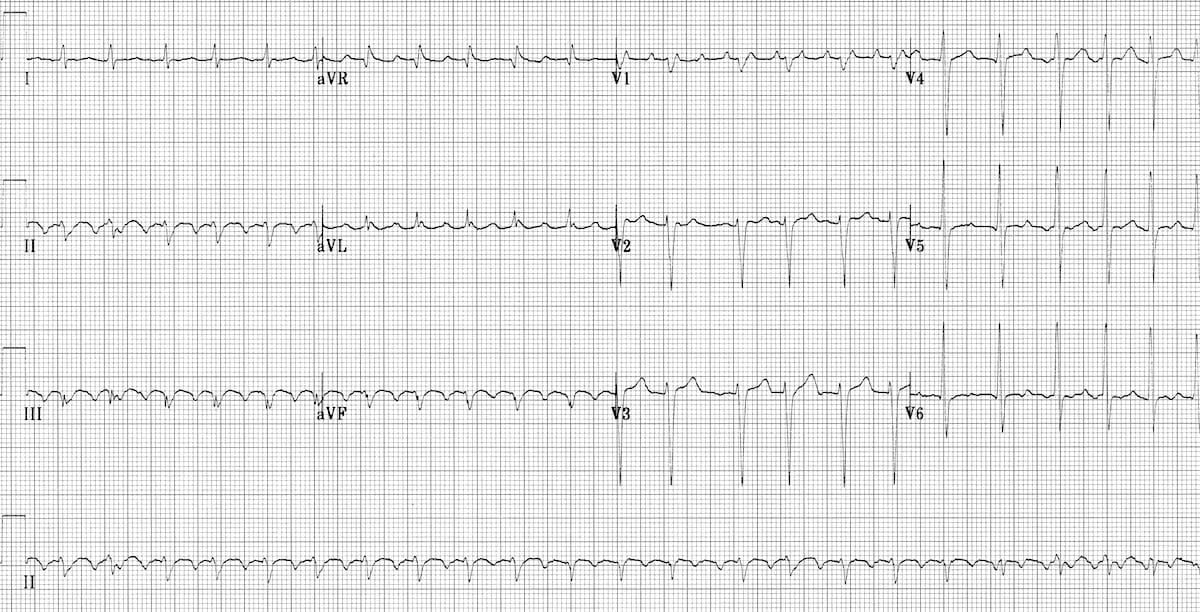
Atypical atrial flutter originates from the left atrium or areas in the right atrium, such as surgical scars, and has a variable appearance on ECG in regards to the flutter waves.In this video we’ll be looking at how to interpret an EKG strip, specifically atrial flutter and atrial fibrillation. This appears as positively-directed flutter waves in the inferior leads. This results in negatively-directed flutter waves in the inferior leads.Īt times, the direction of the circuit can reverse, causing clockwise atrial flutter from the same anatomical location.

Typical atrial flutter rotates counterclockwise in direction, from a reentrant circuit around the tricuspid valve annulus and through the cavo-tricuspid isthmus. Also, atrial flutter can be described as “clockwise” or “counterclockwise” depending on the direction of the circuit. In this situation, giving adenosine will transiently slow the ventricular rate, unmasking the atrial flutter waves and allowing a more definitive diagnosis to be made.Ītrial flutter can described as “typical” (type I) or “atypical” (type II) based on the anatomic location from which it originates. When the heart rate is significantly elevated - that is, greater than 150 bpm - it is often difficult to determine atrial flutter from atrial fibrillation, atrial tachycardia or atrioventricular nodal reentrant tachycardia, or AVNRT. This results in the rhythm becoming “irregularly irregular.” There are only two other rhythms that are commonly irregularly irregular, including atrial fibrillation and multifocal atrial tachycardia, or MAT. In this situation, there may be three P waves to one QRS complex, then a quick change to two P waves to one QRS complex, and so on any combination of P waves to QRS complexes can occur. The regularity of the QRS complexes frequently present with atrial flutter helps to distinguish it from atrial fibrillation, though atrial flutter with variable conduction of the P waves can also occur. In this situation, the ventricular (QRS) rate will be exactly 150 bpm and regular.ĬLINICAL PEARL: A narrow complex tachycardia at a ventricular rate of exactly 150 bpm is very commonly atrial flutter. Typically, the atrial rate will be about 300 bpm, and only every other atrial depolarization will be conducted through the AV node. Just as in atrial fibrillation, not all of the P waves are able to conduct through the atrioventricular node, and thus the ventricular rate will not be as fast as the atrial rate.


 0 kommentar(er)
